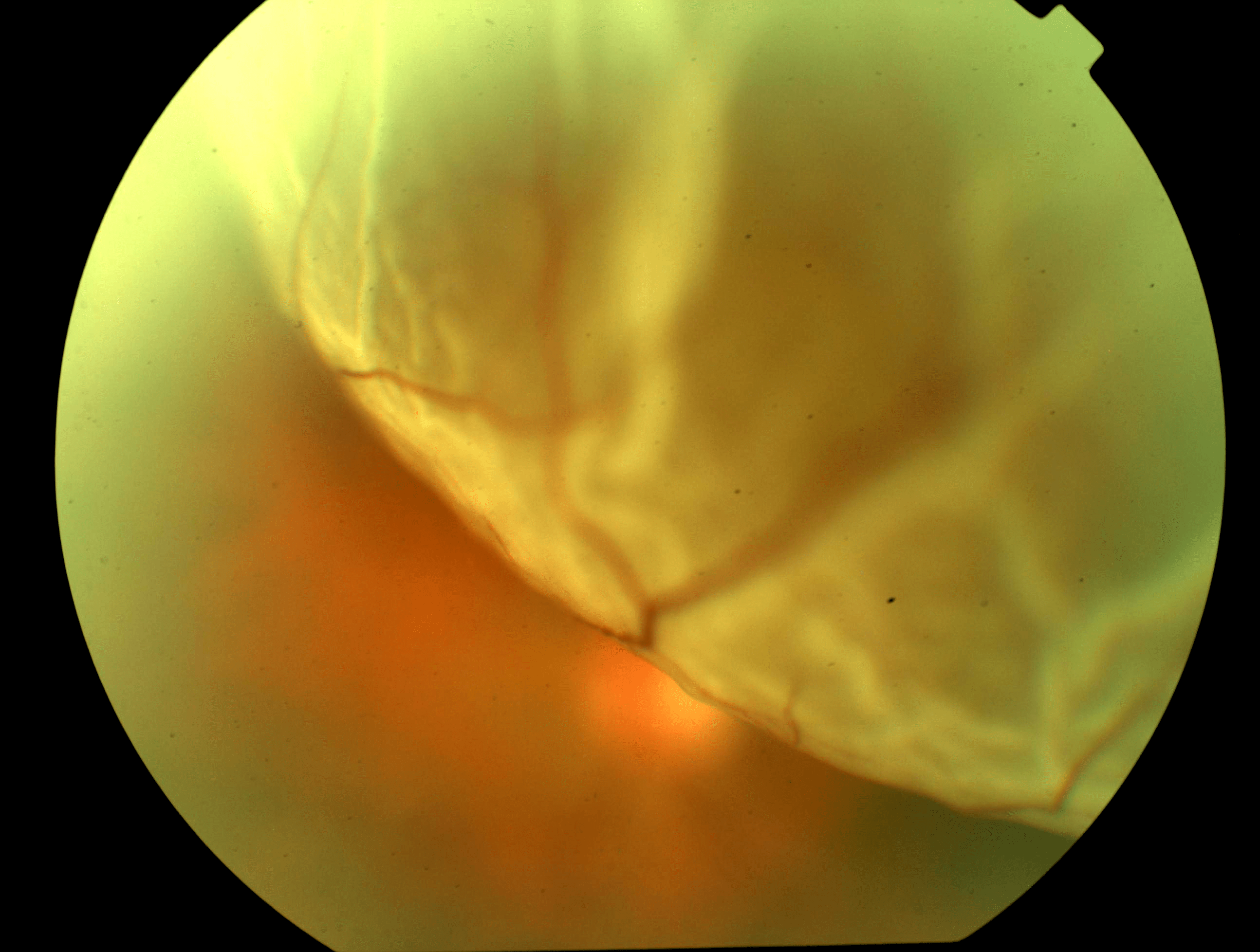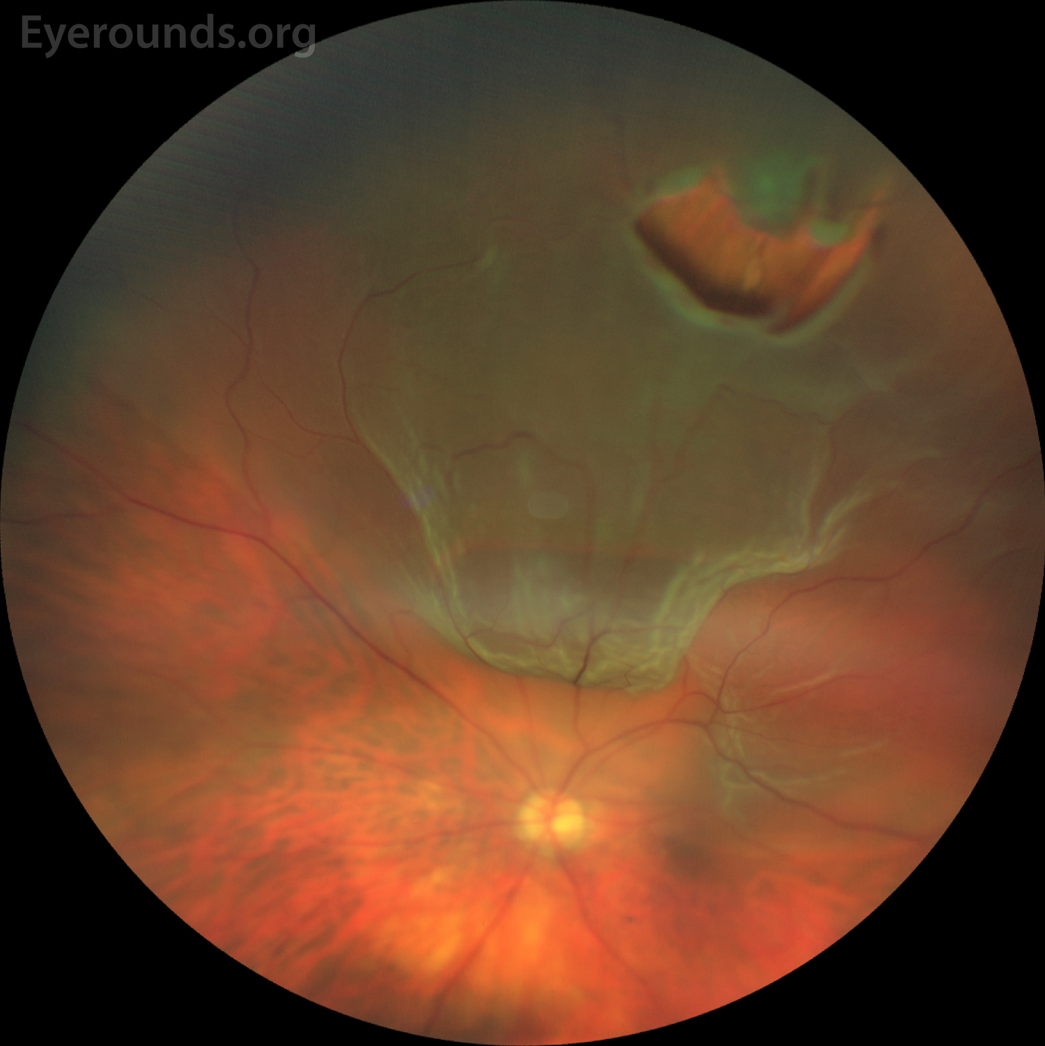

The burns create small scars that fix the tear and help hold your retina in place. Wearing protective eye gear while you participate in any risky activities, such as playing sports, can help reduce your chances of injury. Laser surgery (photocoagulation) With this treatment, your doctor will shine a medical laser inside your eye and make small burns around the tear or hole in your retina. There is no way to prevent aging, which is a major risk factor for getting a detached retina, says VanderBeek. There are no alternative or complementary therapies to fix a detached retina, says VanderBeek. The vitreous may be replaced with a clear fluid. During the surgery, very small openings are made in the eye wall, and most of the vitreous (the gellike fluid that fills the eye) is removed.Īfter that, the surgeon may inject an air or gas bubble into the eye to hold the retina in place and may use a laser or freeze the retina to reattach or repair it. Numbing drops, shots, or general anesthesia are used to make sure there is no pain during the procedure. Vitrectomy A vitrectomy is similar to a pneumatic retinopexy, but the surgery is more involved and is usually performed in a hospital rather than a doctor’s office. If the central retina was detached prior to surgery, vision often improves after a successful reattachment, but some degree of permanent vision loss can occur. In cases when the center of the retina was not detached prior to surgery, postsurgery vision tends to be about the same as preoperative vision. The macula is where you do all your fine seeing - it’s necessary for driving, reading, and seeing fine detail,” says VanderBeek.

“In the central part of your retina, there’s an area called the macula. Retinal detachment repairs are successful in 9 out 10 cases, though sometimes more than one procedure is necessary to successfully reattach the retina. With prompt diagnosis and treatment, the prognosis can be good for retinal detachment. An OCT is a noninvasive test that uses light waves to take cross-section pictures of the retina so that each layer is visible. Browse 30+ retinal detachment stock photos and images available, or search for macular degeneration or pink eye to find more great stock photos and pictures. ComplicationsĮighty-five percent of people with posterior vitreous detachment have no other problems caused by the detachment.If the eye doctor isn’t able to make a diagnosis from the dilated eye exam, an ultrasound or optical coherence tomography scan (OCT) of the eye may be performed to see the exact position of the retina. Photophobia often accompanies albinism (lack of eye pigment), total color deficiency (seeing only in. Light sensitivity also is associated with a detached retina, contact lens irritations, sunburn and refractive surgery. That prompt treatment can lead to better vision-preserving results. Other common causes of photophobia include corneal abrasion, uveitis and a central nervous system disorder such as meningitis. Quick assessment through a dilated eye exam can lead to faster treatment if there is a more serious problem.

The doctor will perform a dilated eye exam, which will widen your pupil and allow the doctor to examine the vitreous and retina. If an eye doctor is not available, go to the emergency room. The symptoms of a PVD often mirror the symptoms of complications such as retinal detachment or a retinal tear.įor this reason, it's important to see an eye doctor quickly if you are having floaters for the first time or if you have more floaters than usual or you have flashes of light, and especially if you have a dark curtain or shadow moving across your field of vision. Although a vitreous detachment is usually harmless, you could go on to develop a sight-threatening complication such as a retinal detachment.


 0 kommentar(er)
